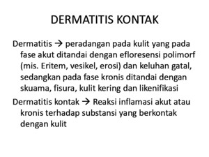
Dermatitis Kontak
There is document - Dermatitis Kontak available here for reading and downloading. Use the download button below or simple online reader.
The file extension - PDF and ranks to the Legal forms category.

342
views
Tags
Related
Comments
Log in to leave a message!
Description
jurnal
Transcripts
Inpatient management of atopic dermatitis dth_1400 249255 S helley D C athcart A my T heos Department of Dermatology, University of Alabama at Birmingham,Birmingham, Alabama ABSTRACT: Atopic dermatitis (AD) is a common chronic inflammatory skin disease that is generally managed on an outpatient basis However, a significant percentage of patients may develop complica-tions severe enough to require inpatient treatment The most common complications of AD that may require hospital admission include erythroderma, eczema herpeticum, and systemic bacterial infec-tion Hospital admission can also be useful for chronic and severe disease that has not responded tostandard therapy or in situations where nonadherence is suspected as the cause of treatment failure Inthese cases, inpatient treatment can offer an opportunity for caretaker education and allow for anobjective evaluation of a patient’s response to a structured treatment plan This article will review theindications for inpatient management of AD and the therapies that can be used to acutely managesevere disease and associated complications KEYWORDS: atopic dermatitis, eczema herpeticum, erythroderma, hospitalized patient, wet wraptherapy Introduction Atopic dermatitis (AD) is a chronic inflammatory skin disease that affects approximately 17% of chil-dren and 1–3% of adults (1) Traditional therapy consists of emollients, mild to moderate potency topical corticosteroids, or topical calcineurininhibitors, along with avoidance of irritants andallergens Although this treatment strategy resultsin significant improvement for most patients, asmall percentage will require more intensivetherapy The most common complications of ADthat may require inpatient management includeerythroderma, eczema herpeticum, and systemicor severe bacterial infection Hospital admissioncan also be useful for chronic and severe diseasethat has not responded to standard therapy or insituations where nonadherence is suspected as thecause of treatment failure In these cases, inpatienttreatment can offer an opportunity for caretakereducationandallowforanobjectiveevaluationofapatient’s response to a structured treatment planThis article will review the indications for inpatientmanagement of AD and the therapies that can beused to acutely manage severe disease and associ-ated complications Erythroderma Acute decompensation of moderate to severe ADcan lead to erythroderma, which is defined as thepresence of erythema and scaling involving greaterthan 80% of the body surface area AD is one of the more common causes of erythroderma, butmanyothercausesexist(Table 1)Regardlessoftheunderlyingdisease,complicationsoferythrodermacan be life-threatening and can include tempera-ture instability, hypoproteinemia, hypovolemia,hypernatremia, and high-output congestive heartfailure Inpatient management of erythroderma isoften necessaryThe first step in the management of a patientpresenting with erythroderma is a thorough Address correspondence and reprint requests to: Amy Theos,MD, Department of Dermatology, University of Alabamaat Birmingham, EFH 414, 1530 3rd Ave S, Birmingham, AL35294-0009, or email: amytheoschsysorg 249 Dermatologic Therapy,Vol 24, 2011, 249–255 Printed in the United States · All rights reserved © 2011 Wiley Periodicals, Inc DERMATOLOGIC THERAPY ISSN 1396-0296 workup to identify the underlying etiology A careful history should be taken at presentation,giving special attention to preceding illnesses,recent fevers, new medications, and relevant pastmedical and family history Physical examinationshould include evaluation of the mucosal surfaces,testingforapositiveNikolskysign(sloughingoftheepidermis with light lateral friction on the skinsurface), and palpation of the lymph nodes Base-line laboratory evaluation should consist of liverfunction tests, chemistry panel, and completeblood count Preexisting dermatitic lesions, severepruritus, lichenification, personal or family history of atopy, elevated serum levels of immunoglobulinE, or eosinophilia can be helpful features thatsupport the diagnosis of AD Helpful clues on his-tological examination include mild to moderateacanthosis and spongiosis, dermal eosinophils,and parakeratosis Although definitive treatment of erythrodermadepends on the underlying cause, all patientsshould be managed for fluid loss, electrolyteabnormalities, and temperature instability (2)Nutritional support is also critical The large body-surface-to-body-mass ratio in children combined with an impaired skin barrier and cutaneousvasodilation leaves them especially vulnerable tohypernatremic dehydration, high-output cardiacfailure and hypothermia (3) Secondary bacterialinfection with Staphylococcal aureus is common inerythrodermic atopic patients and should be rec-ognized and treated promptly Once the cause of erythroderma has been established, proper treat-ment of the underlying condition can be initiatedIn the case of AD, wet wrap therapy can achieverapid improvement Wet wrap therapy Wet wrap treatment is defined as any treatmentconsistingoftheapplicationofatopicalagentwitha double layer of bandages or gauze, first with amoist layer and then a dry second layer The effec-tiveness of wet wrap therapy in AD is thought to bemultifactorialWet wraps have been shown to healthe disrupted skin barrier of AD patients, repairthe lamellar structure of intercellular lipid, anddecrease transepidermal water loss (4) It is alsosuggested that the dressings create a physicalbarrier to scratching, which allows the skin to healin addition to providing a moist environment in which topical medications are more easily absorbed (5) Although most regimens includethe use of a topical corticosteroid, there are alsoreports of improvement with emollient alone A trialbySchnoppet alcomparingwetwraptherapy with either mometasone or a bland emollientshowed significant improvement in both arms of the study; however, the effect was significantly better in the mometasone-treated group (6) Theeffectiveness of steroid-free treatment does,however, lend support to the idea that improvedpenetrationoftopicalmedicationsisonlyonefacetof the effectiveness of wet wrap therapyRegarding the practical details of wet wraptherapy, a basic protocol that includes the featurescommon to most wet wrap regimens is presentedin Table 2 A variation in which the first layer issoaked in warm steroid cream instead of water hasalso been reported to be effective; in this variation,the steroid-soaked wet layer replaces the applica-tion of a topical steroid (7)The steroid of choice as well as the frequency of wet wrap application alsovaries by institution Table 3 shows several topicalsteroids and frequencies of application that havebeen reported effective Regarding the duration of Table 1 Differential diagnosis of common causesof erythroderma DiseaseCharacteristic clinicalfeatures Atopic dermatitis Severe pruritus,lichenification, personal orfamily history of atopy,elevated immunoglobulinE, eosinophiliaSeborrheic dermatitis Infants, greasy yellow scale,diaper area involvedPsoriasis Silvery scale, preexisting psoriatic lesions, nailchanges, + family history of psoriasisStaphylococcal scaldedskin syndromeInfants and young children,superficial blisters, + Nikolsky’s sign, skin foldand perioral accentuation,painfulIchthyosis Congenital, possiblecollodion membraneNetherton syndrome Onset in infancy, sparse hair(trichorrhexis invaginata),failure to thrive, atopy Immunodeficiencies Early onset, alopecia, failureto thrive, recurrentinfectionsDrug History of medication intakeCutaneous T celllymphoma Adults, severe pruritus,keratodermaPityriasis rubra pilaris Keratoderma, islands of sparing, salmon-colorederythrema Cathcart Theos 250 therapy, all studies showed significant improve-ment of AD after 1 week of wet wrap therapy, withless improvement seen in the second week andthereafter(6–10) Therefore, the present authorsrecommend a treatment period of no more than 7daystobalancetheriskofsideeffectswhileachiev-ingthemaximalbenefitavailablewiththistherapyThe major risk of wet wrap therapy is suppres-sion of the hypothalamic-pituitary-adrenal (HPA)axis Several studies have shown that wet wraptherapy can lead to a temporary drop in early morning serum cortisol levels (7,8) However, cor-tisol levels returned to normal within 2 weeks of discontinuing therapy, and no adverse eventsoccurred in either study as a result of this suppres-sionSeveralinvestigatorshaveexaminedtheeffectof dilution of steroid with emollient as a way todecrease the total amount of steroid delivered tothe skin and have found that there was no differ-ence in efficacy between various dilutions of fluti-casone proprionate (see Table 3 for dilutionsstudied) (8,9) Wolkerstorfer et al also examined whether or not HPA axis suppression could beminimized if a more dilute steroid cream wasapplied (9) They found that more HPA axis sup-pression did occur in patients treated with highersteroid concentrations Therefore, increasing thedilution of potent steroids appears to result inlower risk of HPA axis suppression while maintain-ing efficacy of higher steroid concentrationsThe two most common adverse effects associ-ated with wet wrap treatments are poor toleranceof the process and folliculitis likely secondary toocclusion Using creams instead of ointments may reduce the incidence of folliculitis The increasedrisk of bacterial or herpetic infections with wet wrap therapy is controversial but appears to berare Although some studies report an increase inthe incidence of secondary bacterial infection,others actually report a decrease in the coloniza-tionof Saureus ontheskinofADpatientsafterwet wrap treatment (6,8) The present authors suggestthat wet wraps not be applied to any obviously infectedareasuntilproperantimicrobialtreatmenthas been initiated and improvement is seen Eczema herpeticum (EH) EH occurs when skin previously affected by ADbecomes secondarily infected with herpes simplex virus (HSV) EH can lead to serious and even fatalcomplications, especially in the case of dissemi-natedviremiaandmultiorganinvolvement(11,12)For this reason, EH is another complication thatcan require inpatient management DisseminationoftheherpesvirusinpatientswithADisthoughttobearesultoftheimpairedskinbarrier,whichfacili-tates viral invasion and binding to cellular recep-tors (13) Children affected by EH will often havea history of a more severe disease, an earlier onsetof AD, and a higher rate of coexistent food allergy or asthma (14) Elevated serum immunoglobulinE has also been implicated as a risk factor for EH(15) Athough topical corticosteroids have beenimplicated in the past as a potential cause of thiscomplication, further research has refuted thisassertion (15)EH presents as umbilicated vesicles progressing to punched-out crusted ulcers occurring primarily in skin affected by AD (FIG 1) The head and neck are preferentially involved The infection can be aresult of either dissemination of recurrent HSV or aresult of inoculation from an infected person (15)Theskinlesionscanbeextremelypainfulandareatrisk for secondary bacterial infection New lesionstypically appear for the first 7–10 days, and thelesions will typically resolve in 2–6 weeks (16) Theprimaryepisodeisoftenassociatedwithfever,lym-phadenopathy, and malaise Systemic viremia canlead to multiorgan involvement, most commonly the liver, lungs, central nervous system, andadrenal glands The diagnosis can be confirmed with viral changes seen on skin biopsy, direct fluo-rescent antibody (DFA) staining, viral culture, orpolymerase chain reaction for viral DNA on asmear or tissue sample Diagnosis can also bemade performing a Tzanck test The Tzanck testrefers to a procedure in which the base of a freshly unroofed vesicle is scraped and the cells aresmeared onto a glass slide After staining the slide withWright or Giemsa stain, the presence of multi-nuclear viral cells or balloon cells with typical viral Table 2 Wet wrap protocol General wet wrap protocol• Using tubular bandages, cut the appropriate size tocover the arms, trunk, legs, and (if needed) a mask for the faceYou will need enough for two layers• Gently bathe skin with lukewarm water for 5–10minutes Pat dry gently• Apply steroid cream or ointment to the skin• Soak the first layer of bandages in lukewarm water, wring out, and apply to skin wrapping theextremities and trunk• Apply second layer of dry bandages• Leave in place at least 2 hours and repeat every 12hours Children may sleep overnight in dressings if tolerated Inpatient management of atopic dermatitis 251
Recommended